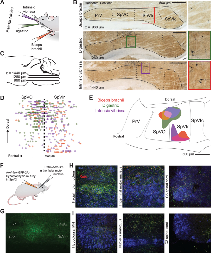
Figure 4. SpVO and SpVIr have two distinct premotor neuron clusters.

(A) Schematic illustration of muscle injections of Pseudorabies-GFP into the biceps brachii, digastric, and intrinsic vibrissa muscles.
(B,C) Example of horizontal sections from an injection in the biceps brachii, digastric, and intrinsic vibrissa muscles, oriented as in panel C. Green fluorescent protein was converted to dark product and sections were counterstained with cytochrome oxidase.
(D) Sagittal reconstruction of all premotor labeled neurons in SpVO and SpVIr.
(E) Two clusters found using the 50 % densest labeling of digastric and biceps premotor neurons and 42 % of intrinsic premotor neurons in SpV are overlaid on a sagittal atlas section (Paxinos and Franklin, 2008), see Methods. (SpVO cluster has 14 biceps, 88 digastric, and 57 intrinsic vibrissa premotor neurons. SpVIr cluster has 33 biceps, one digastric, and 34 intrinsic vibrissa neurons. Total premotor neurons was 94 biceps, 178 digastric, and 218 intrinsic vibrissa premotor neurons across nine mice).
(F) Schematic illustration of the injection scheme for a Cre-dependent AAV with somatic GFP and synaptophysin-mRuby into SpVO and a retrograde AAV-Cre into the facial motor nucleus.
(G)Image of GFP labeling at the anterograde injection site in SpVO.
(H) Images of axon and terminal labeling in the retrograde injection site of the facial motor nucleus (left) and of collateral axons and terminals in the trigeminal motor nucleus (middle) and the lower cervical spinal cord (right).
(I) Example section of terminal labeling in the hypoglossal motor nucleus (left) and lack of labeling in the region of motor neurons in the nucleus ambiguus (middle) and the upper cervical spinal cord (right).
