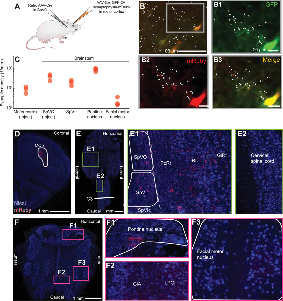
Figure 6. Motor cortex collaterals broadly target brainstem premotor nuclei.
(A) Injection scheme for collateral labeling with a retro AAV-Cre injected in SpVO and AAV-flex-GFP-2A-Synaptophysin-mRuby in motor cortex.
(B) Pyramidal neurons (red and green) and labeled axons (green) and synapses (red and green) labeled by AAV-flex-mGFP-2A-synaptophysin-mRuby. White dots (left top image) indicate counted synapses with arrows (three enlarged images) showing co-labeling at synapses. Motor cortex injection coordinates: Bregma + 3.0, 2.0 lateral.
(C) Quantification of synaptic density in motor cortex, pontine nucleus, facial motor nucleus, SpVO, and SpVIr (50,473 boutons and 135 cells across 3 mice). See white outlines of motor cortex injection, SpVO, SpVIr, the facial motor nucleus, and the pontine nucleus (panels D-F) for areas used for density calculation.
(D) Large view of motor cortex injection site described in panel A with white outline used for density calculation.
(E) Retrograde injection into SpVO shown with collaterals found in SpVIr (panel E1), the medullary reticular formation (panel E1), and the cervical spinal cord (panel E2). White outlines for SpVO and SpVIr used in density calculation.
(F) Collaterals of SpVO-projecting motor cortex neurons in the pontine nucleus (panel F1), the alpha part of the giganticellular reticular formation (panel F2), the lateral paragiganticellular reticular formation (panel F2), and the facial motor nucleus (panel F3).

