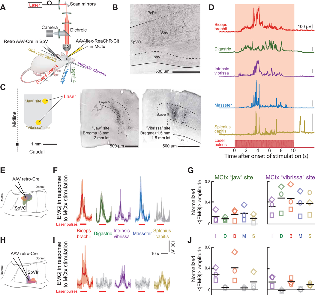
Figure 7. SpVO- and SpVIr-projecting motor cortex neurons drive different networks that activate muscles reflecting their respective premotor clusters.
(A) Schematic illustrating the injection scheme as well as the later used red laser and recorded EMGs in the bicep brachii, digastric, intrinsic vibrissa, masseter, and splenius capitis muscles.
(B) Axon terminals in SpVIr, an example of a retrograde AAV-Cre injection site.
(C) Schematic of injection site and corresponding laser size (left). Cortex injection “jaw” site coordinates: bregma + 3.0, 2.0 lateral with. Cortex injection “vibrissa” site coordinates: bregma + 1.5, 1.5 lateral. Virus-labeled pyramidal neurons from a jaw MCtx and a vibrissa MCtx injection (middle and right respectively).
(D) Envelopes of the EMG signal for the biceps brachii, digastric, intrinsic vibrissa, masseter, and splenius capitis muscles for a single trial. Note that the intertrial interval was 50 s.
(E-G) Activation of EMGs using AAV retro-Cre targeted to the SpVO premotor cluster (panel E). Stimulus-triggered averages from stimulation the MCtx “jaw” site (panel F) and averages across mice (panel G) from jaw motor cortex (left) and vibrissa motor cortex (right) (10 – 20 measurements per muscle per cortex location across three mice). Grey traces in panel F or symbols in panel G show insignificant responses (Student’s t-test, p < 0.05).
(H-J) Activation of EMGs using AAV retro-Cre targeted to the SpVIr premotor cluster. Data are formatted as in panels E to G (10 – 20 measurements per muscle per cortex location across three mice).

