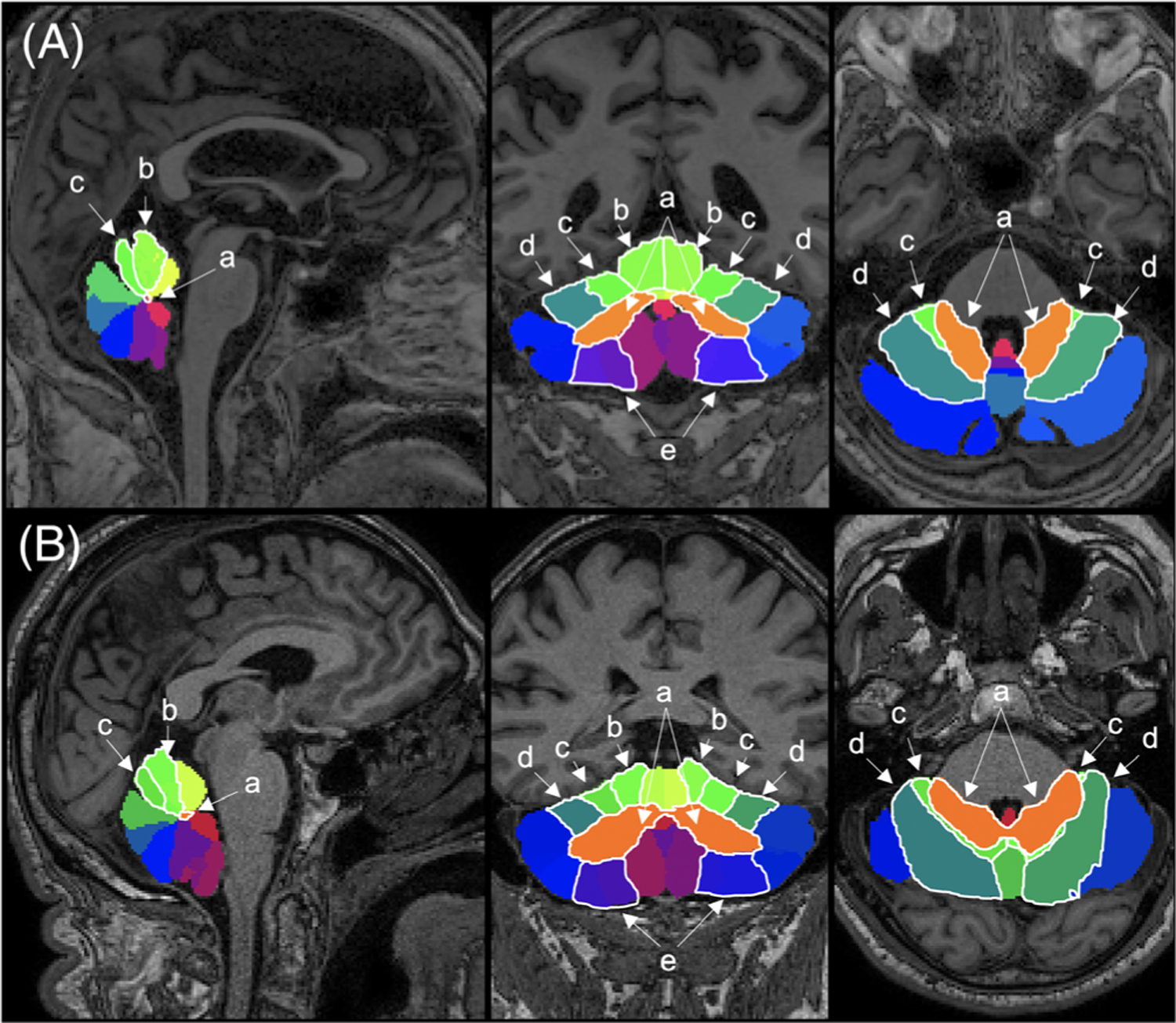
FIG. 1.

Multiatlas cerebellar segmentation. Sagittal (left), coronal (center), and axial views for (A) representative ET patient (male, 68 years) and (B) PD patient (male, 64 years). Specific lobules: (a) deep cerebellar nuclei, (b) lobule IV, (c) lobule V, (d) lobule VI, and (e) lobule VIII are outlined in white. [Color figure can be viewed at wileyonlinelibrary.com]
