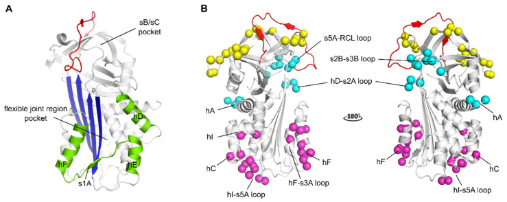
Figure 2.
Localization of binding regions for PAI-1 inhibitors in the structure of active PAI-1. (A) Localization of the binding regions for small molecule PAI-1 inhibitors. The binding pocket in the flexible joint region is aligned by hD, hE, hF and strand 1 (shown in green). The sB/sC pocket is aligned by β-sheet B and C (B), localization of different epitopes of antibodies and antibody fragments as determined by mutagenesis and X-ray crystallographic studies. The epitopes of Abs that prevent the interaction between PAI-1 and PAs comprise residues that are indicated by yellow spheres (exosites for PAs on PAI-1) and residues in the reactive center loop (RCL) (shown in red). The epitopes of switching Abs are indicated by magenta spheres and comprise either residues located in hF and the loop connecting hF and s3A or residues located in the loop connecting hI and s5A, hC and hI. The epitopes of latency-inducing Abs are indicated by cyan spheres and comprise in hA or residues at the top part of the PAI-1 molecule in the hD-s2A loop, the s2B-s3B loop, and the s5A-RCL loop. All panels have been generated using the structure of active PAI-1 (PDB ID 1DB2).

