Figure 4.
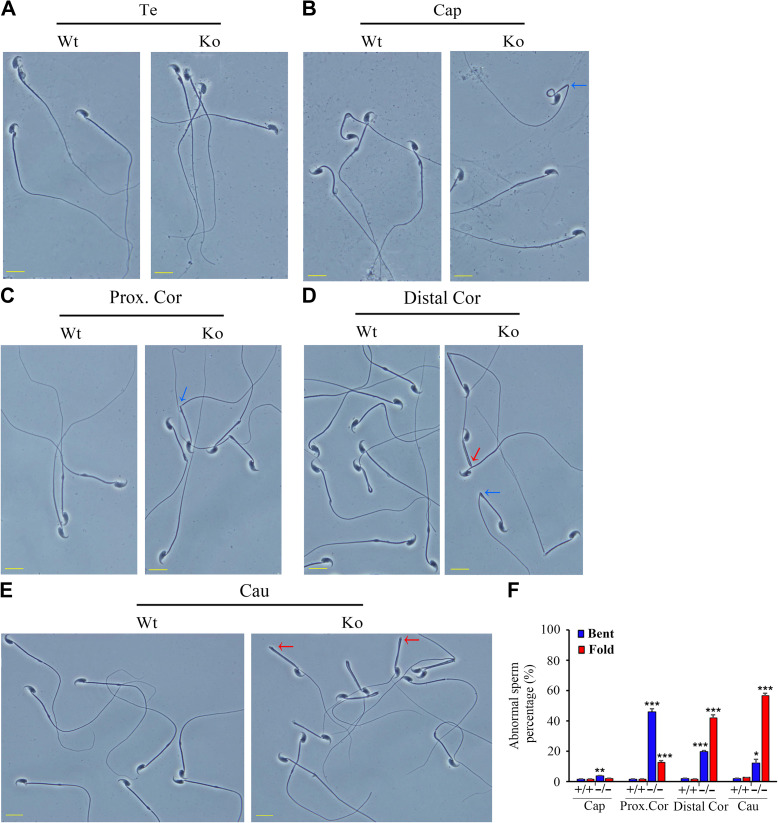
Morphological defects of Emc10−/− spermatozoa. (A−E) Phase contrast images of spermatozoa collected from the testis (A), caput (B), proximal corpus (C), distal corpus epididymidis (D), and cauda epididymidis (E) from wild-type (Wt) and Emc10-null (Ko) mice. Scale bar, 10 μm. (F) Quantification analysis of sperm with abnormal structure in different parts of the epididymis from Wt and Ko mice (n = 3). Data are presented as mean ± SEM, *P < 0.05, **P < 0.01, and ***P < 0.001 compared with respective controls. Te, testis; Cap, caput; Prox.Cor, proximal corpus; Distal.Cor, distal corpus; Cau, cauda epididymidis. Blue arrow, spermatozoa with bent tails; red arrow, spermatozoa with fold tails.

