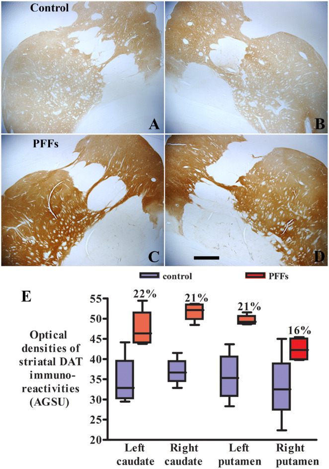
Figure 2.
Microscopy images show DAT staining in monkeys receiving the sham surgery (control; A and B) and α-syn PFFs (C and D). The striatal DAT staining was light and homogenous in the monkey with sham surgery (A and B). Intensive DAT staining displayed in both caudate nuclei and putamen in monkeys receiving PFFs (C and D). Scale bar in D = 830 μm (applies to all). Quantitative optical density measurement (E) revealed further that the optical density of DAT staining was higher in striatum of monkeys receiving the α-syn PFFs than sham surgery (E; P < 0.01). AGSU = arbitrary greyscale units.

