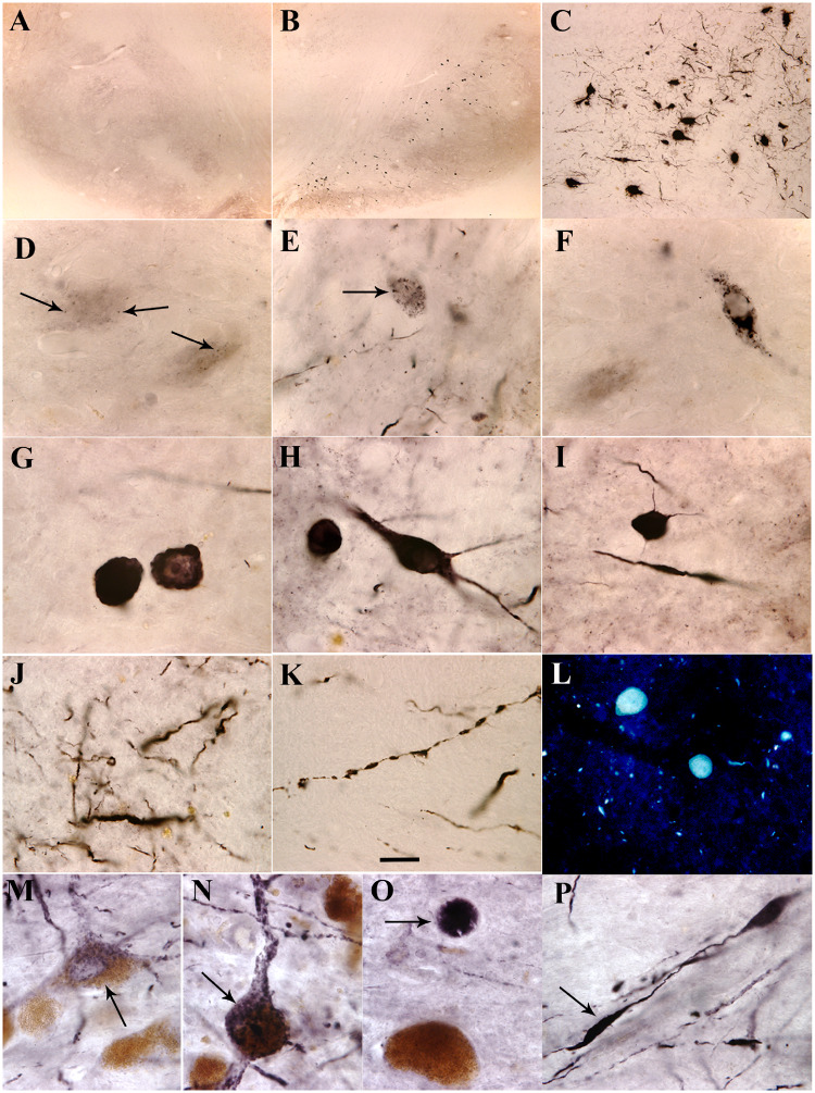
Figure 4.
Microscopy images of substantia nigra. Shown are α-syn phosphorylated on Ser129 (p-S129-α-syn) immunoreactivity (A–K and M–P) and thioflavin S staining (L) from monkeys received α-syn PFFs (B–L), sham surgery (A) and patient with Parkinson’s disease (M–P). Abundant p-S129-α-syn immunoreactive cells were distributed in substantia nigra pars compacta (B and C). These cells exhibited several morphological features: granules (arrows, D–F) with cytoplasmic staining, typical Lewy body with absent cytoplasm (G), and whole cells filled with p-S129-α-syn-immunoreactive products (H and I). Fragmental p-S129-α-syn-immunoreactive neuropil threads were observed in substantia nigra (J and K). Thioflavin S-positive inclusions were detected in substantia nigra (L). These pathological characteristics observed from monkeys received α-syn PFFs were similar to the Parkinson’s disease; p-S129-α-syn immunoreactive products displayed granules (arrow, M), whole cell shape (arrow, N), Lewy body-like (arrow, O), and fragmental fibre (arrow, P). Scale bar in K = 20 μm and applies to D–P; 100 μm for C; and 500 μm for A and B.

