Figure 5.

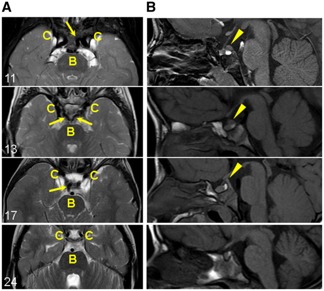
Persistent trigeminal artery and prominent posterior clinoid process in patients with C-terminal truncating MN1 variants. (A) Carotid and basilar arteries (axial view): Persistent trigeminal artery flow-voids (dark signal) connecting the carotid (C) and basilar (B) artery flow-voids (arrows). The persistent trigeminal arteries are unilateral in Individuals 11 and 17, and bilateral in Individual 13. Individual 24 with an early truncating variant does not have persistent trigeminal arteries (shown for comparison). (B) Prominent posterior clinoid process (sagittal view): abnormal tissue just superior to the posterior pituitary bright spot and continuous with the posterior clinoid process (arrowheads). Individual 24 without the abnormal tissue is shown for comparison.
