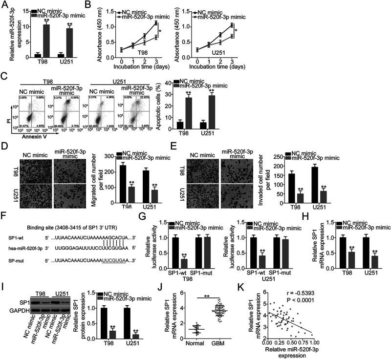
Figure 3.
miR-520f-3p exerts tumor-suppressing actions in GBM and directly targets SP1. (A) miR-520f-3p levels in T98 and U251 cells transfected with miR-520f-3p mimic or NC mimic were determined by qRT-PCR. (B, C) Proliferation and apoptosis in T98 and U251 cells after overexpression of miR-520f-3p were analyzed via CCK-8 assay and flow cytometric analysis. (D, E) Transwell cell migration and invasion assays were conducted to detect the migration and invasion of T98 and U251 cells after miR-520f-3p upregulation. (F) The predicted wild-type and mutant miR-520f-3p binding sites in the 3′-untranslated region (3′-UTR) of SP1. (G) The relationship between miR-520f-3p and SP1 was analyzed by detecting the luciferase activity of SP1-wt or SP1-mut after cotransfection with miR-520f-3p mimic or NC mimic. (H, I) qRT-PCR and Western blotting were conducted to measure SP1 mRNA and protein expression in miR-520f-3p-overexpressed T98 and U251 cells. (J) SP1 mRNA expression in 59 GBM tissues and 19 normal brain tissues was analyzed by qRT-PCR. (K) The correlation between SP1 mRNA and miR-520f-3p expression in the 59 GBM tissues was evaluated by Pearson’s correlation coefficient (r = −0.5393, p < 0.0001). *p < 0.05 and **p < 0.01.

