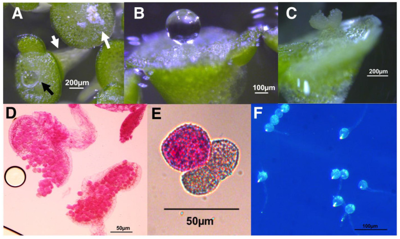
Figure 6.
W. microscopica 2005 flowering and pollen analysis (A) Aerial view of mature W. microscopica 2005 fronds showing the pseudoroot (white arrowhead) extending from the ventral surface of a frond that is asexually reproducing a daughter frond at the same time it has produced a pistil. The pistil emerged from a furrow on the dorsal surface and has secreted stigmatic fluid (black arrow). A dehiscing anther has emerged from the furrow of a different frond (white arrow). (B) Side view of mature pistil with secreted fluid. (C) Side view of a dehiscing bilobed anther. Morphology is as described in Sree et al. [10]. Ruptured anthers releasing pollen (D) and pollen grains (E) stained with modified Alexander’s stain indicating that W. microscopica 2005 is producing viable pollen in culture. (F) 28% of W. microscopica 2005 pollen produced pollen tubes.

