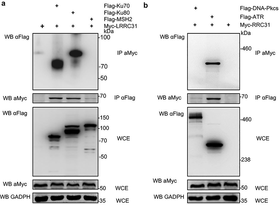
Extended Data Fig. 4. Characterization of the interaction of LRRC31 with Ku70, Ku80 and MSH2 (a) and with DNA-PKC and ATR (b).
HEK293 cells were co-transfected with Myc-tagged LRRC31 and Flag-tagged Ku70, Ku80, MSH2, DNA-PKcs, or ATR. Cell lysates were prepared and immunoprecipitated with anti-Flag or anti-Myc antibody. The precipitated proteins were then separated using 10% (a) and 6% gel (b) SDS-PAGE, and probed with anti-Myc or anti-Flag antibody. WCE, whole cell extract. Two biologically independent experiments were performed with similar results obtained. Unprocessed immunoblots are shown in Source Data Extended Data Fig. 4.

