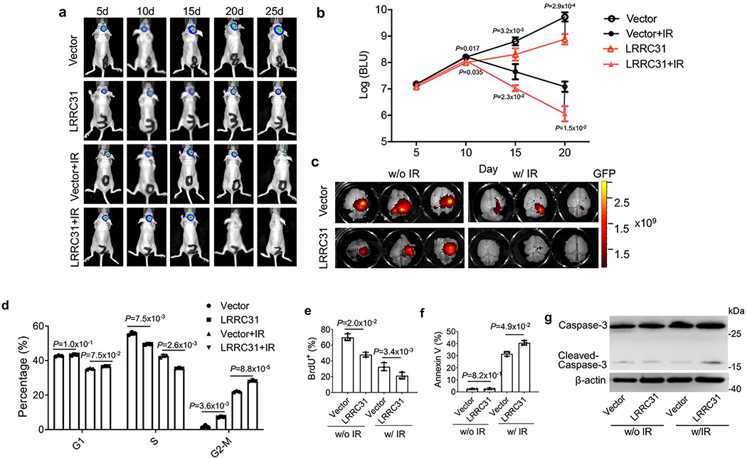
Extended Data Fig. 2. Characterize of LRRC31 for its effects on tumor development in vivo, and on cell cycle, proliferation, and apoptosis in vitro.
Characterize of LRRC31 for its effects on tumor development in vivo, and on cell cycle, proliferation, and apoptosis in vitro. a, b, Representative images of tumors in the brain imaged by IVIS (a) and semi-quantification of the bioluminescence signal (b) in mice received intracranial inoculation of the indicated engineered cells with and without irradiation treatment (5Gyⅹ2). Data show the mean ± s.d. (n=5 animals). c, Ex vivo imaging the brains isolated from mice received the indicated treatment. d-g, Characterization of the effects of LRRC31 overexpression on cell cycle determined by flow cytometry (d), proliferation determined based on Brdu staining (e), apoptosis determined based Annexin V staining (f), and Caspase-3 cleavage determined based on WB analysis (g) in the indicated cells with and without irradiation at 6 Gy. For d-f, data show the mean ± s.d. (n=3 biologically independent experiments). Statistical analysis was performed using the two-tailed, unpaired Student’s t-test. Blot is representative of two biologically independent experiments with similar results obtained. Unprocessed immunoblots are shown in Source Data Extended Data Fig. 2.

