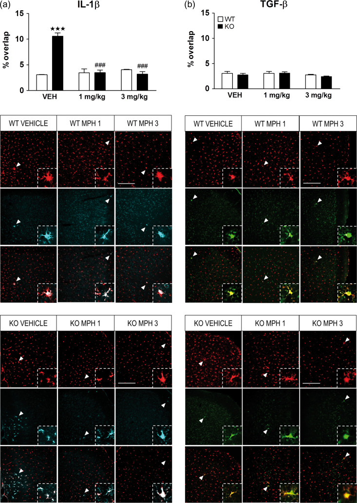
Figure 8.
Chronic treatment with methylphenidate (MPH) restores the enhanced microglial phenotype of IL-1β in the medial prefrontal cortex (mPFC). WT (n = 4) and KO (n = 4) mice were treated chronically for 21 days with vehicle, 1 and 3 mg/kg of MPH and the state of microglia was characterized by double label immunofluorescence of Iba1 with interleukin one beta (IL-1β) a proinflammatory marker (a), and Iba1 with transforming growth factor beta (TGF-β) an anti-inflammatory marker (b). The Y-axis shows the coexpression of microglial cells expressing the pro- or anti-inflammatory marker as a percentage of overlap. The panels on the bottom show representative images of Iba1 positive cells (red) with IL-1β (cyan) or TGF-β (green). White arrowheads represent Co-expression of microglia with IL-1β (left panels) or with TGF-β (right panels) (zoomed in on inset). Scale bar = 200 μm. Images were taken at ×20. The data are mean + SEM cells per mm2. ***P < 0.001 versus WT group with same treatment; ###P < 0.001 versus same genotype treated with vehicle (2-way ANOVA followed by Bonferroni’s post hoc analysis). See Supplementary Table S5 for statistical values.

