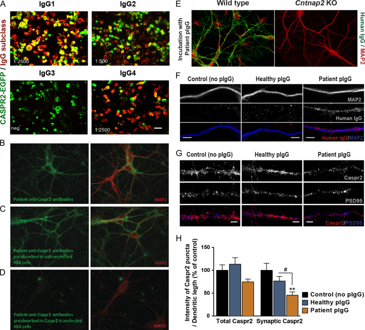
Figure 3.
CASPR2 autoantibodies, predominantly of the IgG1 and IgG4 subclasses, fail to bind to Cntnap2 KO hippocampal neurons and significantly alter the expression of Caspr2 in cortical dendrites. (A) Immunolabeling of human CASPR2-EGFP-transfected HEK293 cells (green) incubated for 1 h with the plasma of a patient with anti-CASPR2 encephalitis. Human immunoglobulins (IgGs; red) are immunolabelled with anti-human IgG antibodies specific for each IgG subclass. End-point titration for each subclass is given in the lower left corner of each panel. (B) Immunolabelling of human IgGs (green) and the neuronal marker MAP2 (red) in hippocampal neurons incubated for 1 h with the patient plasma. (C, D) To confirm the specificity of patient IgGs for CASPR2, and test for the presence of autoantibodies against other neuronal protein targets, patient plasma was pre-absorbed in (C) untransfected HEK293 cells, or (D) HEK293 cells expressing human CASPR2, and incubated with hippocampal neurons. Plasma pre-absorbed in CASPR2-expressing cells fails to stain hippocampal neurons. (E) Immunolabelling of human IgGs (green) and the neuronal marker MAP2 (red) in either wild-type or Cntnap2 KO hippocampal neurons incubated for 1 h with IgGs purified from the patient plasma (patient pIgGs). Patient pIgGs bind strongly to the surface of WT, but not of Cntnap2 KO hippocampal neurons. (F) Immunolabelling of human IgGs (red) and the dendritic neuronal marker MAP2 (blue) in cortical neurons incubated for 7 h with IgGs purified from either the patient plasma (patient pIgGs) or from healthy control (healthy pIgGs). Scale bars = 5 μm. (G) Immunolabelling of Caspr2 clusters in cortical neurons incubated for 7 h with healthy or patient pIgGs. Scale bars = 5 μm. (H) Fluorescence intensity of total and PSD95-co-localised synaptic clusters of Caspr2 was quantified. N = 3, n ≥ 30 cells; Kruskal–Wallis test, Dunn’s post hoc test, **P < 0.01 compared with control, #P < 0.05 relative to healthy pIgG. Results are presented as mean ± SEM.

