Figure 6.
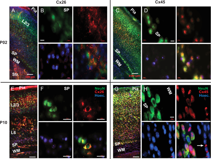
Connexin 26 and 45 immunolabeling. (A) P2 mouse. Composite image (Zeiss Mosaic) comprised of multiple tiles under 20× objective. Double immunolabeling of Cx26 (red) and NeuN (green) in P2 animals, showing weak expression of Cx26 in SP neurons at this age. (B) 100× objective. Colocalization of NeuN (green) and Cx26 (red) in the SP neurons at P2 age. (C) P2 mouse. Composite image comprised of multiple tiles under 20X objective. Double immunolabeling with Cx45 (red) and NeuN (green) showing expression of Cx45 in all cortical layers and the SP zone at P2 age. (D) 100× objective. Colocalization of NeuN (green) and Cx45 (red) in the SP neurons at P2 age. (E) P10 mouse. Composite image comprised of multiple tiles under a 20× objective. At P10 age, Cx26 (red) expression increases in layers 2, 3, 5 and in SP zone, compared with age P2. (F) Images obtained with 100× lens show colocalization of NeuN (green) and Cx26 (red) in the SP neurons at P10. (G) P10 mouse. Composite image comprised of multiple tiles under 20× objective. At P10, Cx45 (red) expression increases throughout the cortex and SP zone, compared with P2 mice. (H) 100× objective. Colocalization of NeuN (green) and Cx45 (red) in the SP neurons, at P10. Cx45 also expresses in non-neuronal cells of the white matter (arrow). Scale bars in A, C, E, and G = 100 μm; in B, D, F, and H = 20 μm. SP = subplate; WM = white matter. Str. = Striatum; Hoechst nuclei stain (blue).

