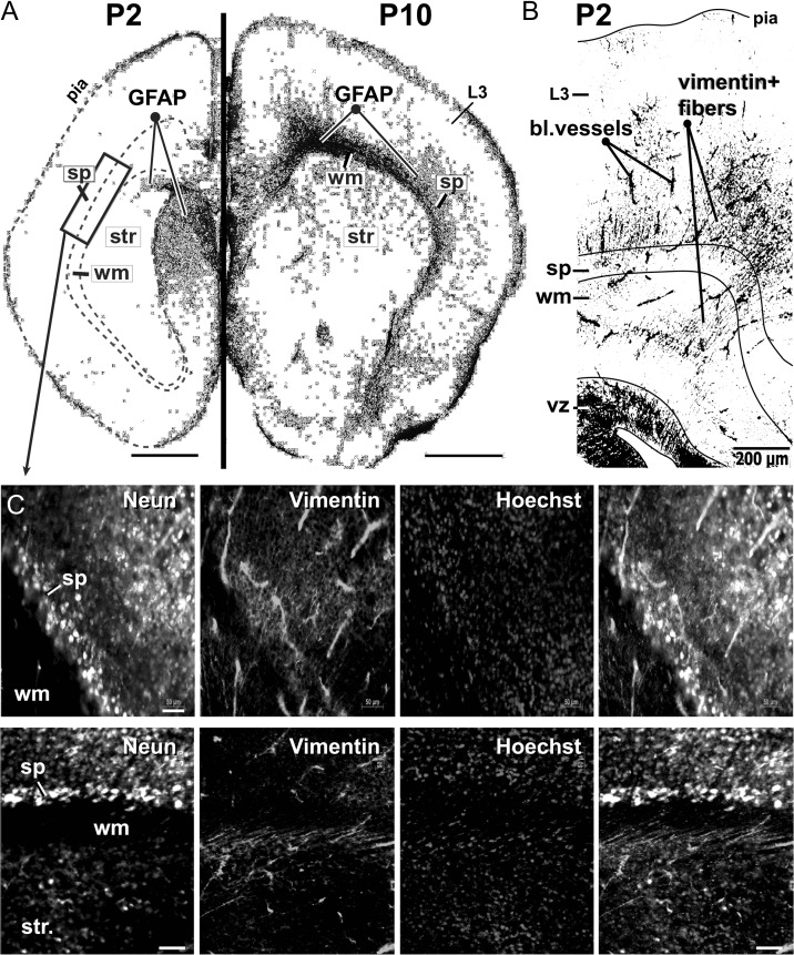
Figure 7.
GFAP and vimentin in the SP zone. (A) Coronal sections of P2 (left) and P10 mouse brain (right), immunolabeled with GFAP (astrocyte marker—black). Each section is a composite of multiple tiles under a 10× objective lens. Images are inverted and equalized. Scale bar = 1 mm. Note that, GFAP is largely absent in P2 cerebral cortex. Rectangle indicates the cortical area used for the acquisition of images at higher magnifications, such as those shown in C. (B) Coronal section of a P2 mouse brain, immunolabeled with Vimentin (black). Scale bar = 200 μm. (C) Subplate zone immunolabeled with NeuN, Vimentin and Hoechst stain. The fourth image is a merge. A high density of vimentin-positive fibers resides inside the SP zone. Scale bar = 50 μm. SP = subplate; WM = white matter; Str = striatum.

