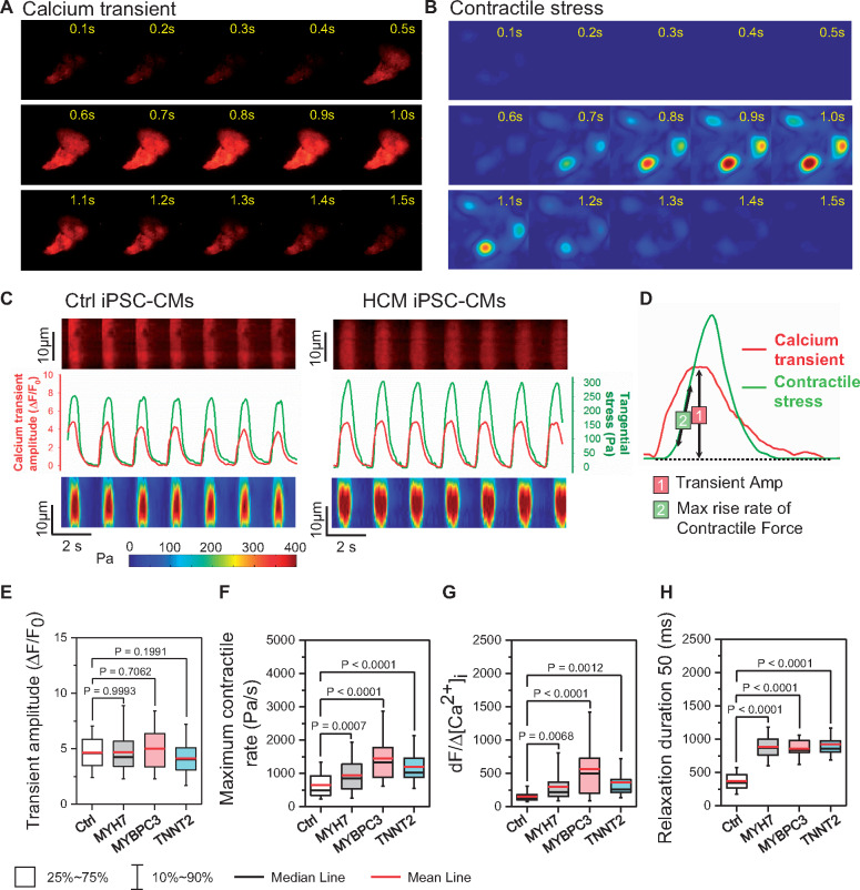
Figure 3.
Functional imaging indicates enhanced risk of diastolic dysfunction in hypertrophic cardiomyopathy (HCM) induced pluripotent stem cell-derived cardiomyocytes (iPSC-CMs). (A–C) Representative traces of simultaneous recording of Ca2+ transient and contractile stress. (D) Measurement of Ca2+ and contraction parameters from a single beating episode. (E–H) HCM iPSC-CMs showed unchanged Ca2+ transient amplitude (E), faster contractile rate (F), increased risk of DD as indicated by Ca2+ sensitivity index (G), and prolonged relaxation duration (H) compared with Ctrl iPSC-CMs by one-way ANOVA (Tukey method). N = 77, 190, 192, and 64 cells in Ctrl, MYH7, MYBPC3, and TNNT2 groups, respectively. For each group, data were generated from two different iPSC lines and three batches of differentiation.

