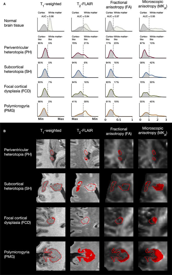
FIGURE 5.
A, Intensity distributions in normal brain and in malformations of cortical development (MCD). In the T1-weighted, T2-fluid-attenuated inversion recovery (FLAIR), and fractional anisotropy (FA) contrasts, the MCD appeared primarily cortex-like, as indicated by the receiver operating characteristic (ROC)-derived thresholds for differentiation of normal cortex and white matter (dashed lines). The exceptions were that polymicrogyria (PMG) exhibited a large portion of white matter-like voxels in T2-FLAIR and that all MCD featured FA distributions with somewhat higher values than in the normal cortex. The microscopic diffusion anisotropy (MKA), however, revealed a mixed cortex-like and white matter-like content within all MCD except periventricular heterotopia (PH), with substantial portions of voxels exhibiting MKA values far into the white matter range. The distributions were averaged across the regions of interest (ROIs) of normal cortex and white matter and all lesion ROIs of each MCD type. The area under the curve (AUC) indicates the area under the curve from the ROC analyses. T1-weighted and T2-FLAIR intensities are normalized, and FA and MKA values are dimensionless. T2-FLAIR intensities are displayed from highest (Max) to lowest (Min). FCD, focal cortical dysplasia; SH, subcortical heterotopia. B, Classifications of tissue as white matter-like (red voxels) in one lesion of each MCD type. T2-FLAIR indicated very high portions of white matter-like voxels in some PMG. FA indicated white matter-like voxels near the border toward white matter of some lesions, whereas the MKA indicated white matter-like voxels also in the deeper parts

