
FIG 1.
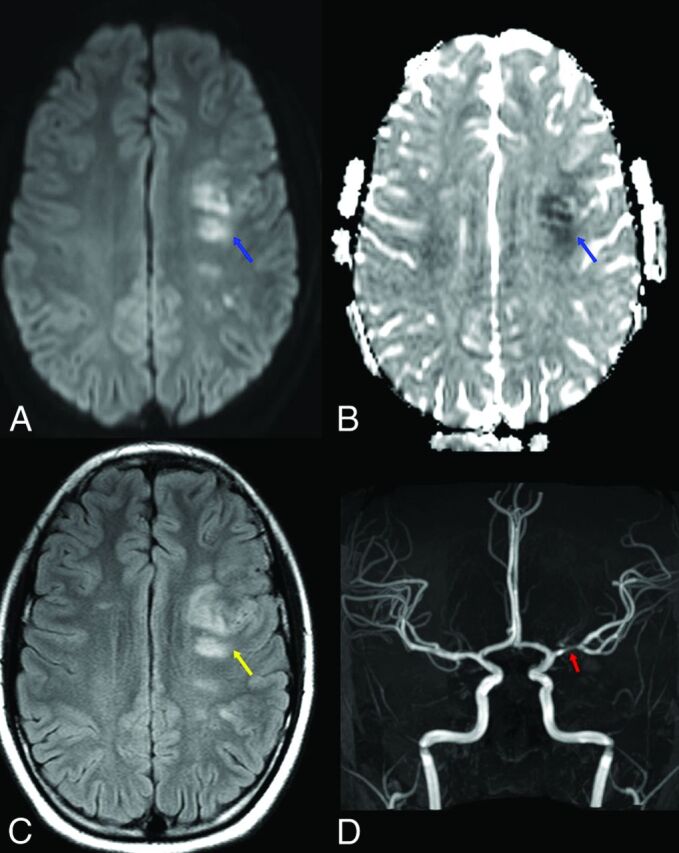
COVID-19-associated left MCA vascular territory acute infarcts in a pediatric patient. Axial diffusion (A), ADC map (B), and FLAIR (C) images demonstrate foci of restricted diffusion (blue arrows) and cytotoxic edema (yellow arrow) within the left middle cerebral artery vascular territory, consistent with acute infarcts. Anterior projection from a TOF-MRA of the head (D) demonstrates a focal segment of moderate stenosis within the left M1 middle cerebral artery (red arrow).