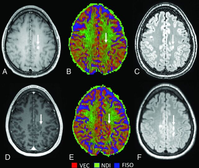
FIG 3.
Lesion type 2 NODDI color map8 graphically representing NDI (green) or VEC (red) compartment prevalence in voxels within a focal WM lesion (white arrows) at baseline (A–C) and at follow-up (after 15 months) (D–F). The green within the lesion is less represented at follow-up after the disappearance of the enhancement, with a relative increase of the red voxels. FLAIR sequences (C and F) show a moderate reduction in size with time. VEC and NDI are fractions in each voxel: If NDI decreases, VEC increases and vice versa. FISO indicates isotropic volume fraction.

