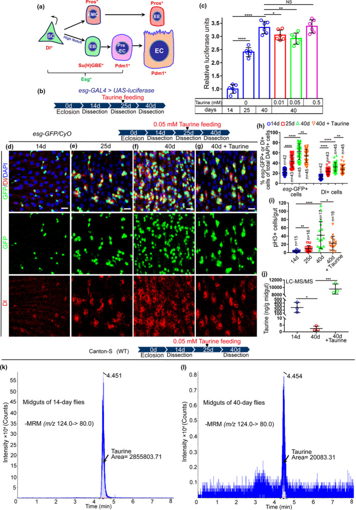
FIGURE 1.

Taurine supplementation represses ISC hyperproliferation in aged Drosophila. (a) Schematic diagram of cell types and markers in the Drosophila midgut. (b) A representative schematic of the Drosophila “esg > luciferase” reporter system. (c) Quantification of luciferase activity of midguts of 14‐, 25‐, and 40‐day flies with or without taurine supplementation. Error bars show the standard deviation (SD) of six independent experiments. (d–g) Midguts were dissected at 14, 25, 40, and 40 days with taurine treatment and stained with Dl (red; ISC marker), and esg‐GFP (green; EB and ISC marker). (h, i) The average numbers of esg‐GFP+, Dl+, and pH3+ cells are significantly lower in 40‐day midguts after taurine supplementation. (j) Quantification of the content of taurine in midguts of 14‐day, 40‐day, and 40‐day flies fed with taurine using LC‐ESI‐MS/MS. Error bars indicate the SD of three independent experiments. (k, i) LC‐MS chromatograms of taurine in midguts of 14‐day (k) and 40‐day (i) flies. Data information: Scale bars represent 10 µm. DAPI‐stained nuclei are shown in blue. Error bars represent SDs. Student's t tests, *p < 0.05, **p < 0.01, ***p < 0.001, ****p < 0.0001. non‐significance (NS) represents p > 0.05. See also Figure S1
