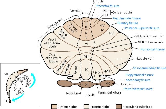
Fig 1.
Unfolded view of the cerebellar cortex showing the lobes, lobules (by name on the right and number on the left), and main fissures (blue font). The hemispheric lobules are designated with the prefix H followed by the Roman numeral indicating their corresponding vermian lobules. Adapted from Haines DE. Fundamental Neuroscience for Basic and Clinical Applications. 4th ed. Philadelphia: Elsevier/Saunders; 2013 with permission of Elsevier.30

