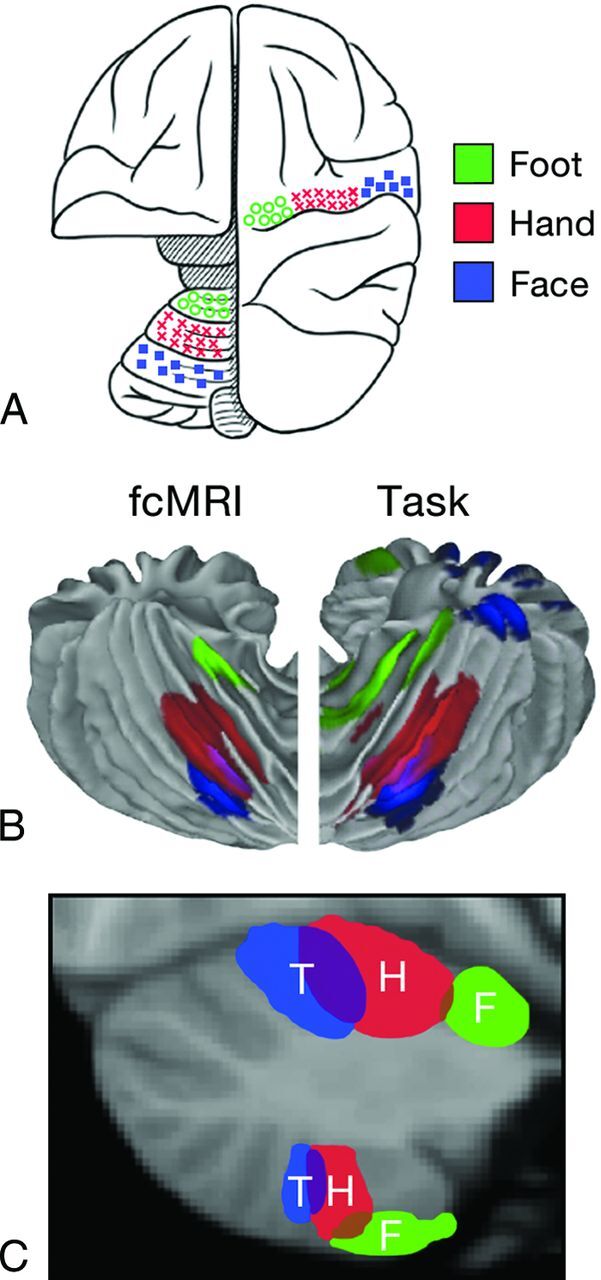
Fig 6.

A, Schematic demonstration of the cerebral and cerebellar functional locations of the foot (green), hand (red), and face (blue) in the monkey. B, Cerebellar locations of the foot (green), hand (red), and tongue (blue) in humans measured by fMRI. “fcMRI” refers to results based on functional connectivity studies. “Task” refers to results from task-based fMRI studies. C, Cerebellar locations of foot (F, green), hand (H, red), and tongue (T, blue) representations in humans from fcMRI studies displayed on a parasagittal image. Note the mirror image representation of the somatomotor functions with the primary or dominant location in the anterior lobe of the cerebellum. Adapted from Buckner RL. The cerebellum and cognitive function: 25 years of insight from anatomy and neuroimaging. Neuron 2013;80:807–15 with permission of Elsevier.31
