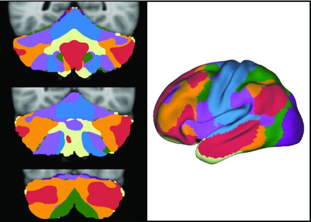
Fig 8.
The 3 images on the left represent multiple coronal sections of the cerebellum with colors representing different cortical functions. The right-sided image is the cerebrum with the colors representing the different functional areas. The somatomotor cortex is blue. This cortex is represented at the more medial aspect of the cerebellum. Most of the human cerebellum, however, is linked to cerebral association networks, including executive networks (orange) and the default network (red). These association networks have multiple cerebellar representations. Adapted from Buckner RL. The cerebellum and cognitive function: 25 years of insight from anatomy and neuroimaging. Neuron 2013;80:807–15 with permission of Elsevier.31

