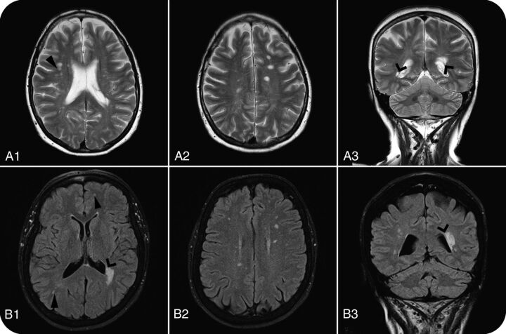
Fig 3.
Brain MR imaging WM anomalies in 61-year-old (A1–A3: T2WI) and 66-year-old (B1–B3: FLAIR) women with RIS. Open arrows show periventricular lesions, and closed arrows show juxtacortical lesions, which, together with >9 lesions, made both patients fulfill the DIS-Barkhof and DIS-Swanton criteria. Columns 1 and 2 are axial sections; column 3 shows coronal sections.

