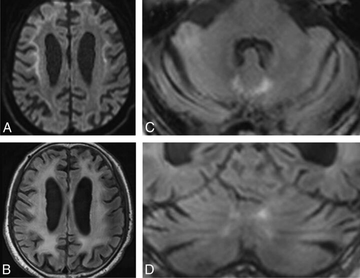
Fig 2.
Patient 4. DWI (A) shows high-intensity signal along the corticomedullary junction. A FLAIR axial image (B) shows diffuse high intensity in the bilateral cerebral hemispheres. FLAIR axial (C) and coronal (D) images show atrophy of the cerebellum and high-intensity signal in the medial part of the cerebellar hemisphere right beside the vermis (the paravermal area).

