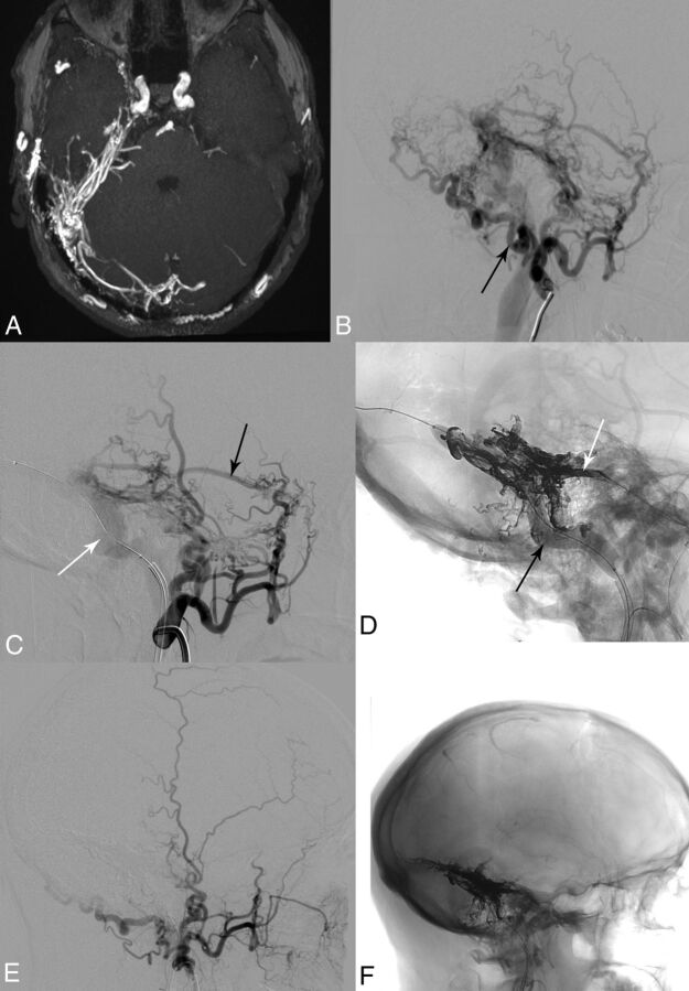
Fig 2.
A 51-year-old man (patient 7) after 3 unsuccessful endovascular treatment attempts 10 years ago showing a progressive Borden I fistula on the right. A, Time-of-flight angiography. B, Right external carotid artery angiogram shows feeding arteries from the middle meningeal artery, occipital artery (black arrow), meningohypophyseal trunk (not visible), posterior auricular artery, artery of the falx cerebelli (not visible), subarcuate artery, and vertebral artery (not visible). C, Right external carotid artery angiogram after positioning of the venous balloon in the right transverse and sigmoid sinuses (8 × 80 mm, Copernic RC; white arrow). Middle meningeal artery (black arrow). D, Fluoroscopic image depicting the inflated venous balloon (black arrow), the arterial double-lumen balloon microcatheter in the middle meningeal artery, partially hidden behind Onyx (4 × 10 mm, Scepter C; white arrow), and the cast of Onyx-18. E, Right external carotid artery angiogram after embolization of the fistula with complete occlusion. F, Fluoroscopic image shows Onyx-18 distribution after complete occlusion of the dural fistula.

