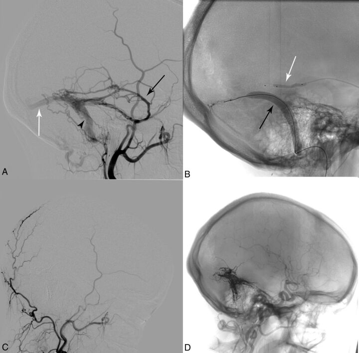
Fig 3.
A 24-year-old woman (patient 1) with a Borden II fistula on the left. A, Baseline left external carotid artery angiogram with feeding arteries from the middle meningeal artery (black arrow), occipital artery, and meningohypophyseal trunk (not visible). Transverse sinus (white arrow); sigmoid sinus (black arrowhead). B, Fluoroscopic image showing an inflated venous balloon (8 × 80 mm Copernic RC; black arrow) and arterial double-lumen balloon catheter in the middle meningeal artery (4 × 10 mm, Scepter C; white arrow). C, Left external carotid artery angiogram (2-month follow-up) after embolization of the fistula showing complete occlusion. D, Fluoroscopic image depicting Onyx-18 distribution after complete occlusion of the dural fistula.

