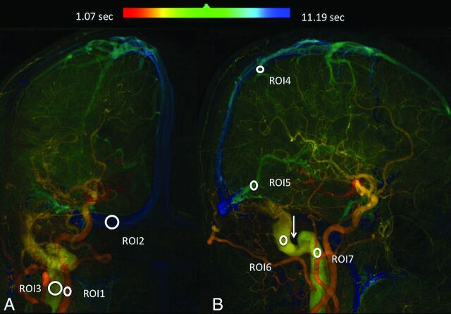
Fig 1.
Quantitative color-coded digital subtraction angiography of the anteroposterior (A) and lateral (B) views of a Cognard type I DAVF. ROI1: internal carotid artery; ROI2: ipsilateral normal transverse sinus; ROI3: internal jugular vein; ROI4: parietal vein; ROI5: vein of Labbé; ROI6: prestenotic segment; ROI7: poststenotic segment. The Arrow indicates the stenotic sinus segment.

