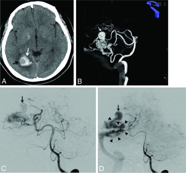
Fig 1.
A, Unenhanced brain CT scan (axial section) in a 40-year-old woman with headache. Right occipital hematoma is seen (white arrow). Note a round hypoattenuated shape surrounded by the hematoma, corresponding to the intranidal aneurysm (black arrow). B, Volume rendering reconstruction from the 3D-RA acquisition through the left vertebral artery, showing a large intranidal aneurysm (white arrow). C and D, Left vertebral DSA in anteroposterior projection at early phase (C) and late phase (D). At early phase, an intranidal aneurysm is seen (C, arrow). On late phase, this nidal aneurysm appears clearly connected to the main draining vein (D, arrowheads), confirming the venous nature of this intranidal aneurysm.

