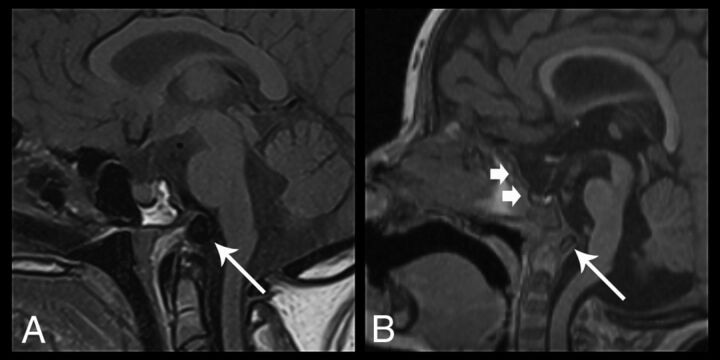
Fig 9.
Sagittal images of skull base dysplasia in 2 different patients. A, Sagittal T1-weighted image demonstrates skull base hypoplasia with dorsal angulation and posterior displacement of a hypoplastic basioccipital ossification center (arrow) and widening of the spheno-occipital synchondrosis. B, Sagittal 3D T1-weighted image shows a J-shaped sella (short arrows) with flattening and elongation of the tuberculum sella. There is also evidence of a dorsally angulated clivus (long arrow), with findings similar to those in A. None of the patients had basilar invagination.

