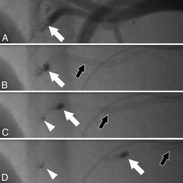
Fig 2.
Fluoroscopic angiography of an ADAPT maneuver. Angiography shows a clot (A, white arrow) in a branch of the axillary artery with a diameter of 1.9 mm. The pressure gradient before and behind the clot is 38 mm Hg, necessitating an aspiration catheter with an inner diameter of at least 1.9F for clot removal, according to our calculations. The clot (A–D: white arrow), which is partially radiopaque, is engaged with a Sofia 5F catheter (B, black arrow). When the catheter is pulled back (C and D, black arrow), the larger portion of the clot can be removed (C and D, white arrow). However, there is fragmentation of the clot, with a small portion of the clot remaining in the vessel (C and D, arrowhead).

