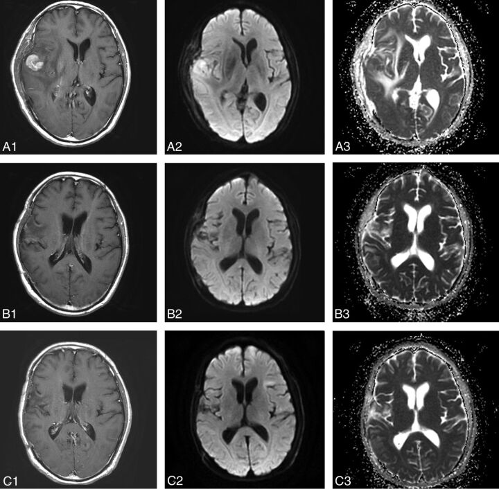
Fig 1.
MR images in a 54-year-old man with diffuse large B-cell PCNSL, belonging to the CR group (A1, A2, A3, before therapy; B1, B2, B3, after 1 cycle of chemotherapy; C1, C2, C3, after 5 cycles of chemotherapy). Contrast-enhanced T1-weighted image shows an apparent enhanced tumor on the right temporal lobe (A1). The tumor shows hyperintense on the DWI (A2, B2). The pretherapeutic ADCmin of the tumor was 668 × 10−6 mm2/s (A3). After 1 cycle of chemotherapy, the size of tumor has decreased significantly (B1, B2) and the ADCmin of the tumor has increased to 1014 × 10−6 mm2/s (B3). After 5 cycles of chemotherapy, the tumor has almost disappeared (C1), and the ADCmin has increased to 1026 × 10−6 mm2/s (C2, C3).

