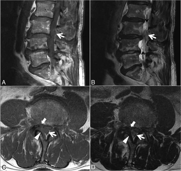
Fig 4.
Case 2. A 77-year-old man with 4 months of low back pain and intermittent radiation into the right-more-than-left lower extremities, as well as weakness and an acute inability to ambulate. Sagittal T1- (A) and T2-weighted (B) and L3 inferior endplate–level axial T1- (C) and T2-weighted (D) images. A heterogeneous predominantly T1-isointense, mildly T2-hyperintense, 1.9-cm maximal-dimension mass (white arrows, A–D) in the dorsal and right lateral epidural fat abutting the right ligamentum flavum (arrowheads, C–D) contributes to severe L3–4 spinal stenosis, with leftward displacement and effacement of the thecal sac. There is no definite connection to the right L3–4 facet joint. The right lateral epidural fat is effaced, and the dorsal mass is contiguous with the dorsal margin of the L3–4 disc (white block arrow in C–D). Differential considerations in the radiologist's interpretation included focal epidural hematoma (particularly given the acute clinical presentation), dorsal migration of a disc fragment, an unusual-appearing synovial cyst or other degenerative cyst, and sequela of the recent epidural injection. At the operation, the dorsal disc herniation was indeed traced back to a large annular defect at the right lateral margin of the L3–4 disc. Also contributing to the L3–4 stenosis are slight retrolisthesis of L3 on L4, a disc bulge, and ligamentum flavum redundancy. Tortuosity of the cauda equina (black arrows, B) is compatible with the high-grade stenosis. An L1–2 left subarticular disc extrusion causing advanced lateral recess narrowing is also present at the superior aspect of the sagittal images (A and B).

