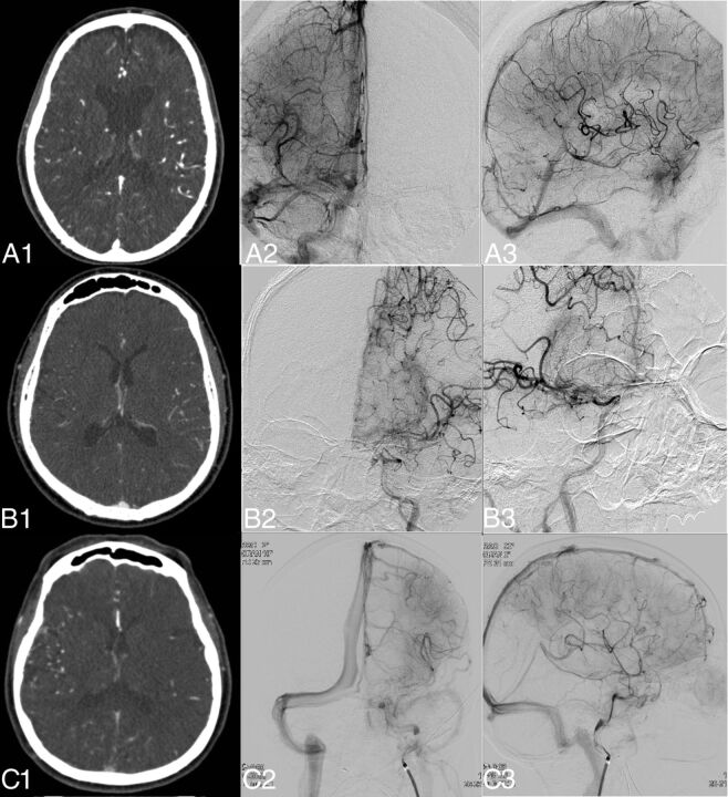
Fig 1.
Examples of DSA- and CTA-based collateral scores in 3 different patients. Images were selected by a maximum amount of contrast in the middle cerebral artery for CTA and adequate opacity in the venous phase for DSA. In the left column, the CTA image is shown (A1–C1); in the middle column, the anteroposterior DSA (A2–C2); and in the right column, the lateral DSA (A3–C3). A1–A3, Patient A with a right-sided M1 occlusion, which DSA assessed as grade 3, and CTA, as grade 3. B1–B3, Patient B with a left-sided M1 occlusion, which DSA assessed as grade 1, and CTA, as grade 3. C1–C3, Patient C with a left-sided M1 occlusion, which DSA assessed as grade 3 collateral flow, and CTA, as grade 1.

