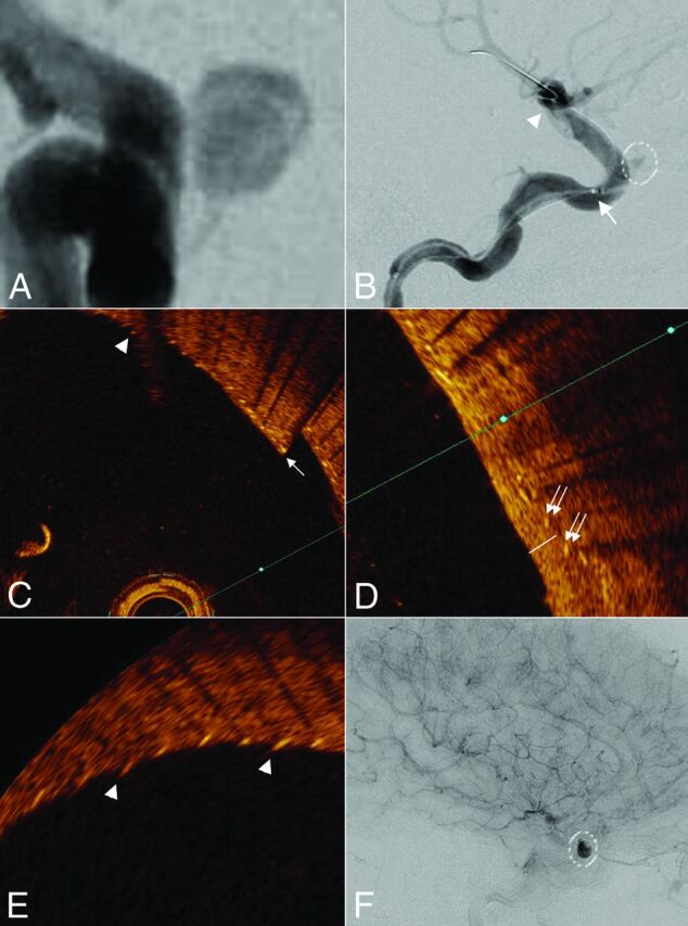
Fig 2.

Case 10. DSA showing collar sign in incompletely occluded paraophthalmic ICA aneurysm after Pipeline embolization (A). Lateral DSA of the ICA showing the aneurysm (white dashed line) and position of the tip (arrowhead) and the beginning of the scanning portion (arrow) of the OCT catheter (B). OCT images showing variable degree of endothelialization (C–E). There are portions where the PED struts lay bare (arrowheads) or are covered with thin endothelium (arrow) or robust neointima (double arrow and white line showing the distance from lumen to PED struts). After treatment with a second PED, there is stasis of contrast within the aneurysm (F).
