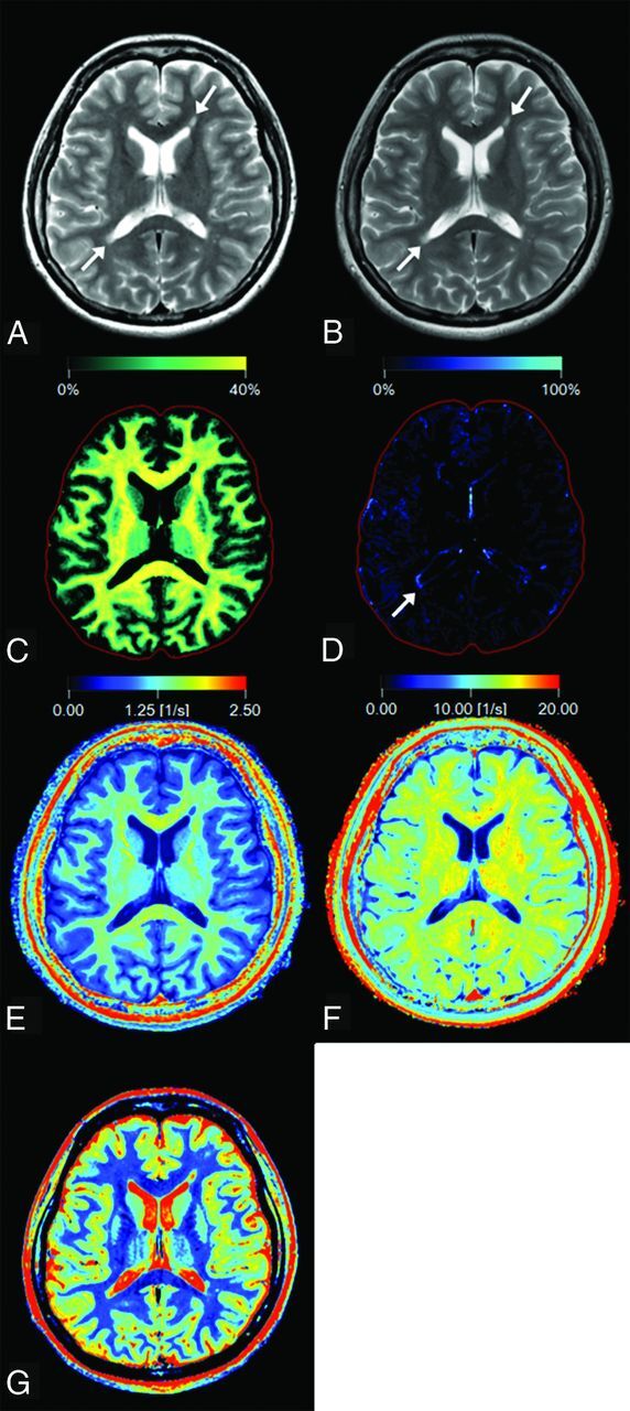
Fig 1.

Representative images of a 27-year-old woman with multiple sclerosis. Panels show a synthetic T2-weighted image (A), a conventional T2-weighted image (B), and maps of myelin partial volume (C), excess parenchymal water partial volume (D), R1 (E), R2 (F), and PD (G). Two plaques are shown by arrows on the T2-weighted images (A and B). On the VEPW map (D), the periphery of the plaque adjacent to the trigone of the right ventricle (arrow) is visible but the one adjacent to the anterior horn of the left ventricle is not. The VEPW of this invisible plaque was very low but still higher than that of NAWM. The red intracranial outline is displayed for visual guidance in tissue images (C and D).
