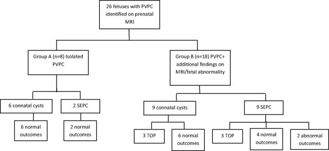
Fig 2.
Flowchart illustrating the study design and outcome. Cases are divided to 2 groups: fetuses in group A had only PVPC on MR imaging, while fetuses on group B had additional findings on MR imaging or fetal abnormality. Fetal abnormality is defined as the presence of fetal infection, chromosomal abnormality, IUGR, abnormal echocardiogram findings, or other fetal malformation. The groups were further subdivided into connatal cysts or subependymal pseudocysts.

