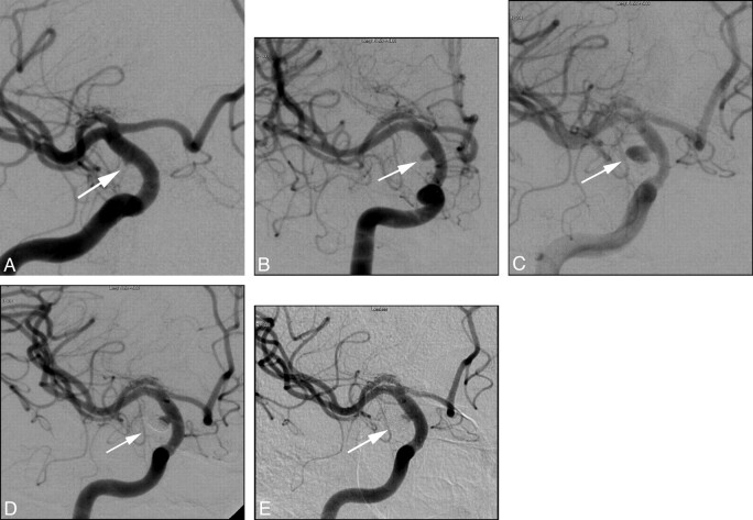
Fig 3.
Right supraclinoid ICA blister aneurysm. Preprocedural DSA (A) shows a shallow outpouching (white arrow) arising from a nonbranching site along the lateral wall of the supraclinoid segment of the right ICA, consistent with a blister aneurysm. One-month follow-up DSA (B) following endovascular stent placement again shows the blister aneurysm (white arrow), now increased in size and more round in shape. The patient subsequently underwent adjunct coil embolization with a single coil. Subsequent 3-month follow-up DSA (C) again shows the blister aneurysm, again increased in size and more round in shape. The patient subsequently underwent adjunct coil embolization with multiple coils. Subsequent 6-month follow-up DSA (D) again shows the blister aneurysm (white arrow), with recurrence at the aneurysm base. The patient subsequently underwent adjunct coil embolization with multiple coils. Twelve-month follow-up DSA (E) shows wide patency of the stents and minimal residual filling of the aneurysm sac (white arrow), with no filling of the coil interstices or aneurysm dome.

