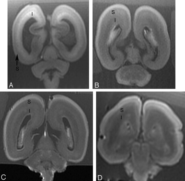
Fig 2.
Postmortem coronal T1-weighted images at (A) 18 weeks, (B) 22 weeks, (C) 23 weeks, and (D) 25 weeks gestational age. At 18 gestational weeks, the intermediate zone (I) is of higher T1 signal intensity and the subplate layer (S) is of lower T1 signal intensity. With increasing gestational age, there is a reduction in the high T1 signal intensity of the intermediate zone and an increase in the signal intensity of the subplate layer. At approximately 22 weeks, the distinction between the intermediate zone and subplate layer is decreased on T1.

