
Fig 2.
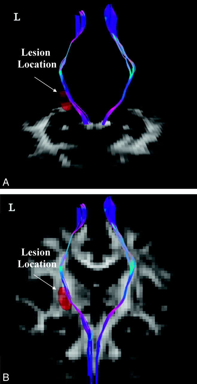
Tractography of bilateral CSTs corresponding to lower extremity motor control in a patient with ischemic stroke at the left PLIC (red area). A, The axial view of the bilateral CSTs passing through the CP; (B) the coronal view of the bilateral CSTs. For fiber direction, red indicates the left-right direction, green indicates the anteroposterior direction, and blue indicates the inferior-superior direction. The background images are GFA maps.