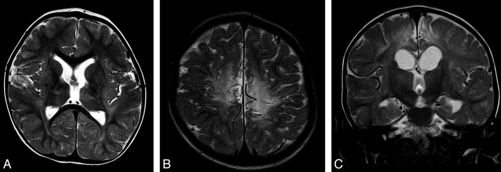
Fig 2.
Typical appearances of HIBD changes in a child with spastic CP following acute profound HIBD. A and B, Axial T2 images show moderate PCWM signal-intensity abnormality and very mild involvement of the putamen and thalamus. C, Coronal T2 image shows no signal-intensity abnormality in the region of the STN.

