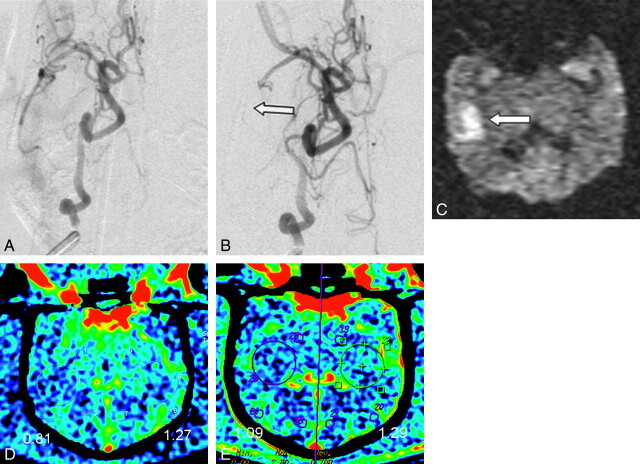
Fig 1.
A−C, DSA with selective ICA injection pre- (A) and postembolization (B) at the origin of the ICA, resulting in an occlusion of the right MCA (arrow in B). C, DWI performed 4 hours later confirms the presence of a right MCA infarct (arrow). D, C-arm CT and PCT (E) demonstrate corresponding decreased CBV as determined both by the color maps and measured values in the respective regions of interest. CBV values are in milliliters per 100 grams of brain tissue.

