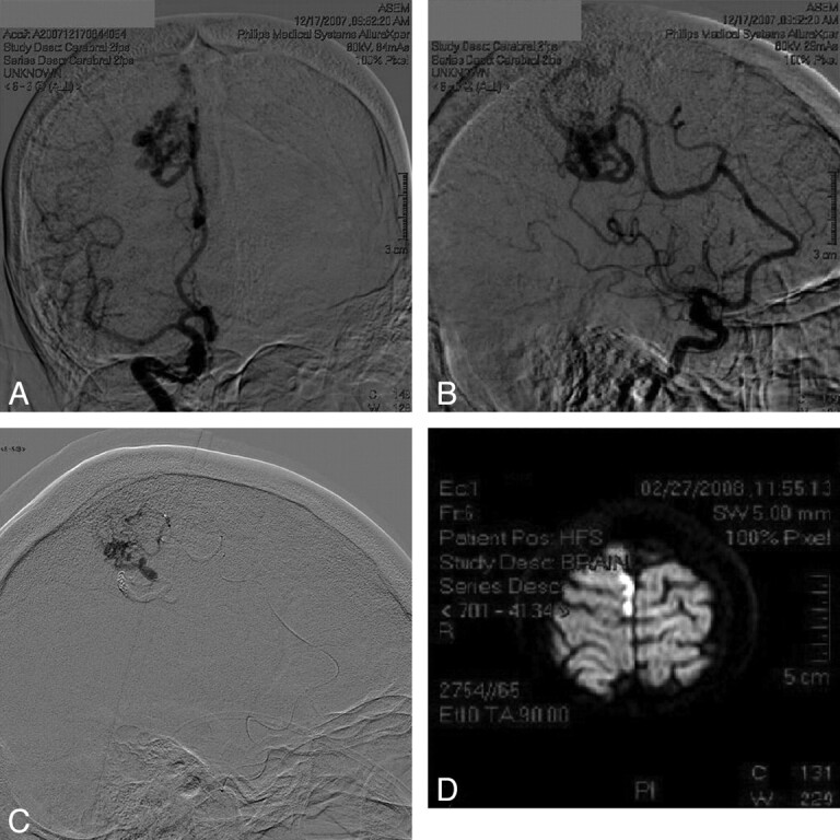
Fig 2.

A 54-year-old woman with a right medial parietal grade 2 AVM who presented with chronic headaches and dizziness. A and B, AP and lateral views of the first diagnostic angiogram. C, Note prior embolization, which required assistance of aneurysm coils due to the fistulous nature of the lesion. Also note the microcatheterization of the AVM nidus through the ACA feeder. After a superselective Wada test with propofol, the patient showed no neurologic changes, yet after embolization, she developed left-leg distal paresis. D, Postoperative diffusion-weighted MR image shows an ischemic insult close to the embolization area. The patient improved to baseline after physical therapy.
