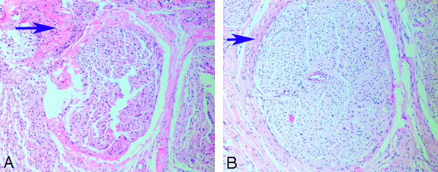
Fig 4.
The arrows demonstrate the thickness of scar tissue of nerve fiber bundles 8 weeks after surgery. A, A rat treated with NGF shows a very thick band of scar tissue surrounding fiber bundles. B, A rat treated with EPO shows very thin bands of scar tissue surrounding fiber bundles (hematoxylin-eosin stain, original magnification ×100).

