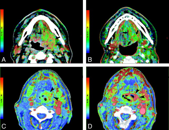
Fig 1.
BF maps at admission (A) and after the completion of 40 Gy (B) of concomitant cisplatin-based chemoradiotherapy in a responder with squamous cell carcinoma of the oropharynx (arrows). Note the significant reduction of the tumor volume at 40 Gy, accompanied by lower BF values. Serial BV imaging at the baseline (C) and after 40 Gy (D) of concomitant chemoradiation in a nonresponder with squamous cell carcinoma of the hypopharynx. The BV maps demonstrate clearly the increase of BV in the tumor site during the follow-up (arrowheads).

