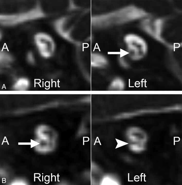
Fig 1.
Two cases of unilateral CND. Magnified oblique sagittal CISS images through the IACs in 2 different patients with ANSD. A, An 8-month-old boy with complete absence of the right cochlear nerve. Compare these findings with the normal-appearing cochlear nerve on the left (arrow). B, A 10-month-old girl with a hypoplastic left cochlear nerve. The left cochlear nerve (arrowhead) is present but is noticeably smaller than the normal-appearing right cochlear nerve (arrow).

