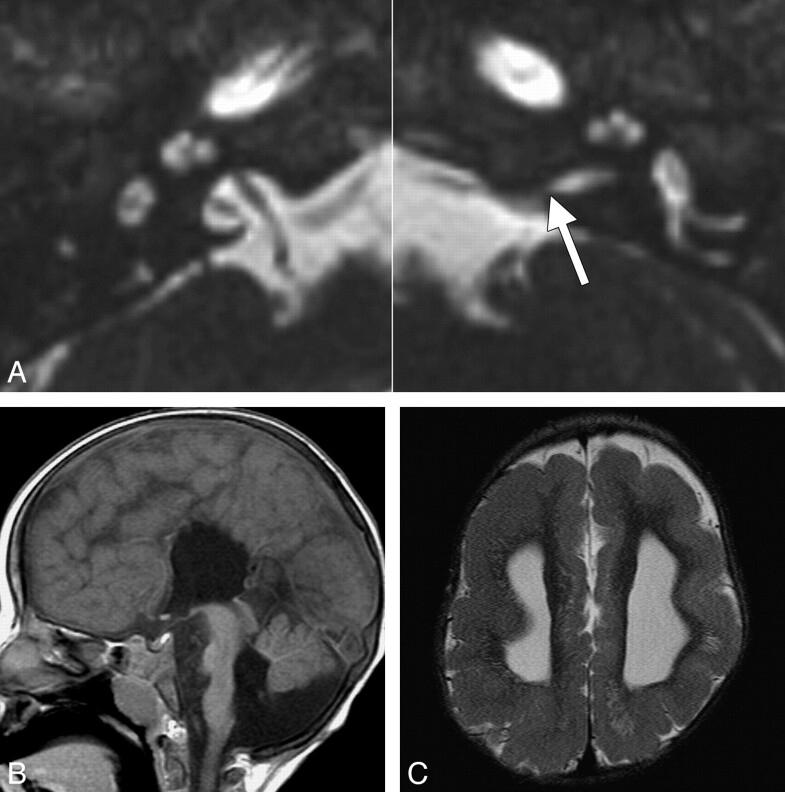
Fig 2.
Inner ear and brain abnormalities in a 2-year-old girl with ANSD and bilateral CND. A, Axial CISS images through the bilateral inner ears and IACs. The left IAC is stenotic, particularly at the level of the porus acousticus (arrow), while the right IAC is normal in caliber. Both cochleae are isolated and dysplastic, with truncated apical turns, and the right modiolus is deficient. B, Midsagittal T1-weighted image demonstrates callosal agenesis, pontine hypoplasia, and inferior vermian hypoplasia. There is a prominent ventral cleft at the level of the pontomedullary junction. C, Axial T2-weighted image through the lateral ventricles demonstrates ventriculomegaly and diffuse pachygyria with relatively few sulci.

