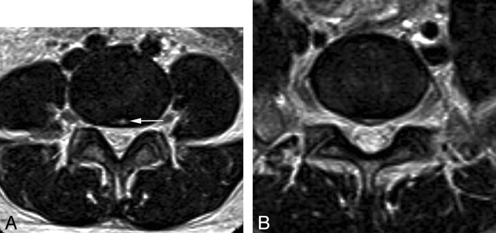
Fig 3.
Annular tear and disk herniation at the central zone. MR image of a 33-year-old woman with LBP. A, On a T2 axial image at the L4–5 disk level, an annular tear at the central zone is seen as the focal high signal intensity in the central zone (arrow), the so-called “high-intensity zone,” B, On a T2 axial image at the L5–S1 level, disk herniation at the central zone is noted.

